Neurons are arguably one of the most important types of cells in our bodies. Recent trends in neural degenerative diseases have pushed researchers towards developing therapies for these conditions. To do so, scientists can use neuronal cell culture and 3D neural organoids to model disease, evaluate cell therapies, and investigate tissue development. In order to properly conduct these experiments, scientists must understand what makes a high quality neuron culture and how to identify one.
What distinguishes a high quality neuron
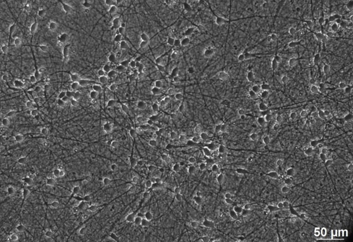
There are several factors that go into identifying the quality of neuronal cultures. Most often, the quality of a neuron is determined by its morphology. Neurons consist of a nucleus and cell body, with dendrites, or processes, and axons that branch from their center. Neurons can be distinguished based on the number, length, and variety of their processes. They communicate with each other through synapses, the point where two or more neurons meet. The larger the synapses, the stronger the electrochemical signal, and the better the quality of the neuron.
How to identify quality neurons
In order to visualize the morphology of cells under a microscope, they first need to be stained with the appropriate cell markers. There are several different accepted methods for staining neurons. Each method can give different information about the neuron (density, number, processes, proteins, etc.), so it is important to understand your needs before selecting a method.
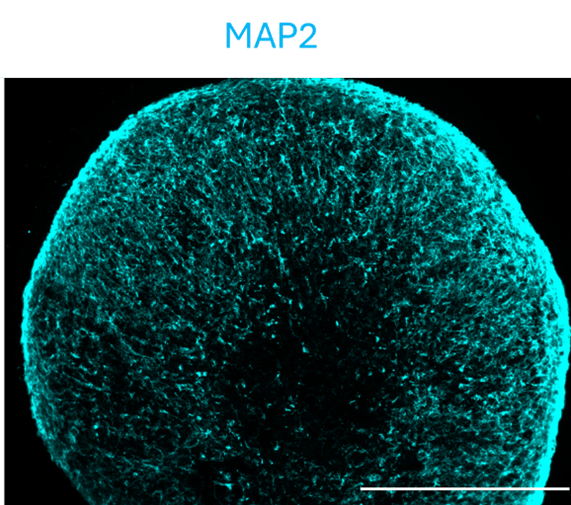
For example, MAP2 is a protein expressed on mature neurons. MAP2 is expressed only on dendrites, making this part of the neuron structure easy to identify once stained. Staining to identify the MAP2 present in a culture of neurons can help determine the maturity of the neurons that are growing. The more MAP2 present, the more mature the neurons. An example of a neural organoid stained with MAP2 is below.
There are other proteins specific to neurons, (for example Synapsin1, which is one of the most neuron-specific proteins) that can be used to identify other parts of the neuron or validate neuron specificity.
How to improve neuronal growth
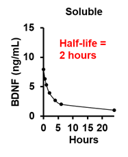
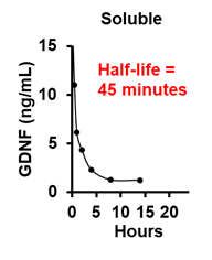
Neuronal growth can be improved by providing neurons with the proper growth at the proper times. Two of the most important growth factors for neuronal growth and development are brain and glial derived neuronal factors (BDNF and GDNF). BDNF promotes neuronal survival, axon elongation and branching, and dendritic spine formation. GDNF enhances axon elongation and branching in dorsal root ganglion. Although these growth factors can be useful individually, their combined breadth and depth is even more advantageous for a wide range of applications.
BDNF and GDNF are often replenished in cultures every 2-3 days. However, BDNF and GDNF have very short half lives in culture (2 hours and 45 minutes, respectively – graphs below). Because of this, cells are starved of these critical growth factors after only a few hours, and they are not replenished again for another 24 hours or more. In order to improve neuronal growth, it is important to maintain the levels of these (and other) growth factors in culture.
Best products for neural generation
One way to control the level of growth factors in culture is by using controlled-release growth factor technology. StemCultures provides StemBeads and DISC Devices that maintain the ideal level of growth factors in culture for up to two weeks.
To product cortical neurons, neural precursor cells were differentiated using Neurobasal base media supplemented with either soluble factor (20 ng/mL) every 2 days or StemBeads growth factor (10 ng/mL) delivery every 4 days for 30 days.
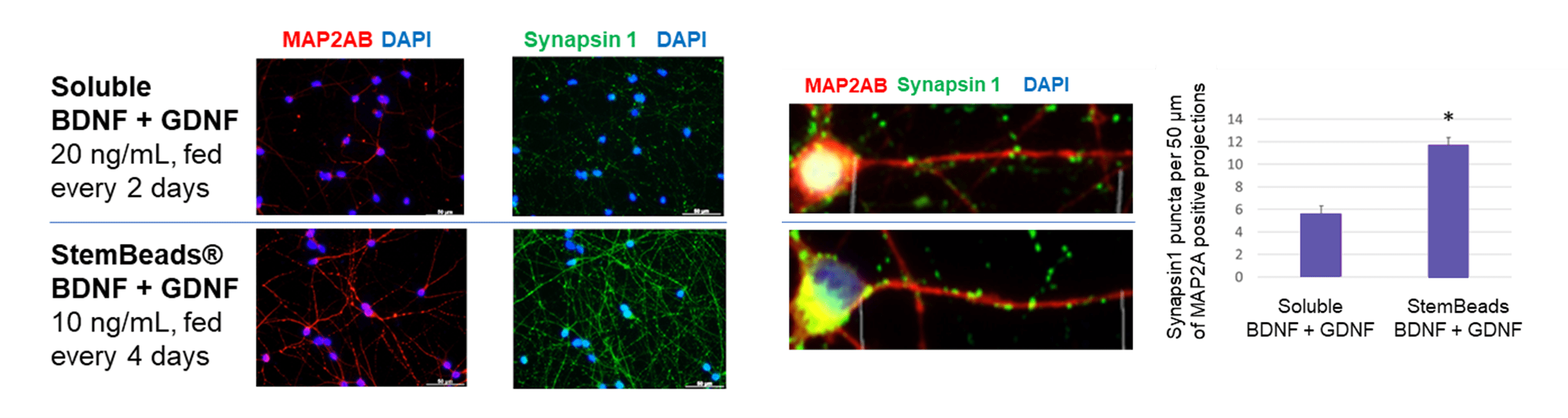
The results showed cells that received BDNF and GDNF with StemBeads controlled-release delivery increased co-localization of Synapsin1 on MAP2A positive neurons. This suggests a greater number of synapses within the culture. StemBeads delivery at a lower concentration and with half as many replenishments produced equivalent or better neurons compared to soluble.
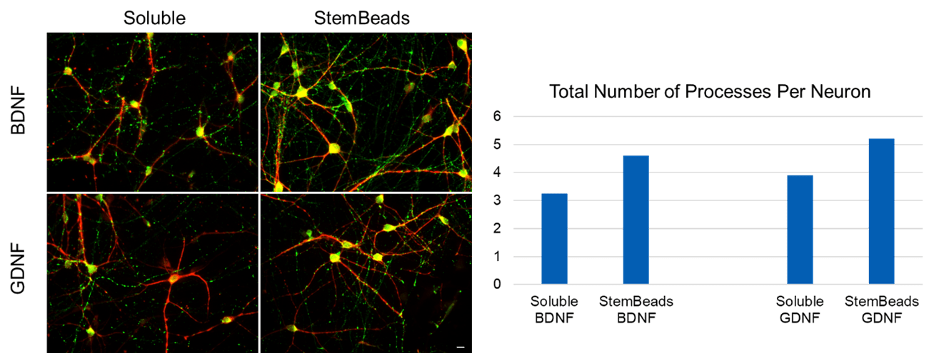
Other experiments show using StemBeads BDNF or GDNF improves the total number of processes per neuron, which is indicative of better neuron quality.
Newly added BDNF and GDNF DISC Devices sustain the release of BDNF and GDNF for up to two weeks in culture. With this technology, feeding is reduced, the protein concentration is reduced, and cells continuously receive the cells they need to be their best.
For more information on these products, or to see how other growth factors can improve your cell cultures, visit our products page or contact us with any questions.
—
Note: The data and images presented in this blog post were generated by members at the Neural Stem Cell Institute and NeuraCell core facility. Opinions and accounts expressed herein are those of the author(s) or interviewee(s). They may not reflect those of StemCultures, its officers, or directors.